Ultrasound is used in both obstetrics (pregnancy) and gynaecology, using high frequency sound waves to create an image.
Ultrasound waves are directed into the abdomen by a transducer placed on the skin or when placed into the vagina. The sound waves are reflected by the interface between organs with different densities. The clarity of the image depends on the frequency of the sound waves and the distance of the transducer from the region being examined. Higher frequencies will allow more detail to be seen but will not travel as far into the body. The closer the transducer can get, the higher the frequency that can be used and the more detail that can be seen.
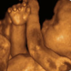
Ultrasound can be performed at different stages of pregnancy:
5-11 weeks Viability/Dating 11-14 weeks Nuchal Translucency Screening 18-20 weeks Fetal Structural Assessment 24-42 weeks Fetal Growth and Welfare Assessment 26 weeks 4-Dimensional