

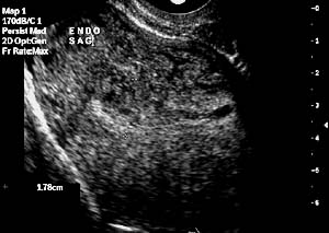
| Sonohysterography is a new technique developed to better image the uterine cavity. It uses an infusion of sterile saline through a soft plastic catheter placed in the cervix in conjunction with transvaginal ultrasound. The saline infusion distends the uterine cavity and provides an excellent contrast to the lining, giving improved visualization of uterine and endometrial pathology. |  |
This technique may also be used to assess the fallopian tubes by demonstrating fluid spill into the pelvis. Colour Doppler imaging demonstrates the movement of ultrasound contrast medium within the tube.
Why is it performed?
The main indications for this procedure include:- Abnormal uterine bleeding both pre and post menopausal
- Investigation of infertility and recurrent miscarriage
- Endometrial assessment for patients on Tamoxifen therapy
- Suggestion of a mass in the endometrial cavity on ultrasound
|
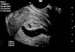 |
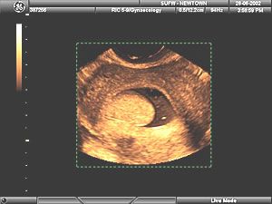
| Sonohysterography - After saline infusion |
This is a simple procedure, able to be performed in the ultrasound room. It does not require sedation or analgesia and is useful before hysteroscopy and dilatation and curettage (D & C).
It allows the presence, nature, size, vascularity and site of attachment of a mass in the uterine cavity to be assessed before definitive surgery.
In a number of cases, it can eliminate the need for further investigation when no significant endometrial pathology can be demonstrated.
This is particularly important in cases of bleeding around the time of menopause where a hormonal disturbance is the most common cause and does not benefit from surgery.
Patients undergoing investigation for infertility have a high incidence of polyps in the uterine cavity which may be responsible for their inability to conceive. This is particularly important in those patients starting on IVF programmes, as these polyps may limit their success.
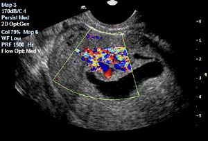 |
| Colour doppler imaging of the endometrial polyp during Sonohysterography |
Patients on long term Tamoxifen therapy for breast cancer, have been shown to develop polyps and thickened uterine linings (endometrial hyperplasia) as well as occasionally developing endometrial cancer.
Although this is a rare complication, it is an important side effects of Tamoxifen.
More commonly, however there changes in the muscular layer just under the uterine lining which is distinguishable from endometrial hyperplasia on sonohysterography but not on standard transvaginal ultrasound.
 |
| 3D image of an endometrial polyp |
